
CASE STUDIES
The Diversity of Shingles

ABOUT
Client: Kelly Cloninger (UIC BVIS)
Software: Pixologic ZBrush, Substance Painter, Keyshot, Adobe Photoshop, Adobe Illustrator
Final Presentation Format: Poster (print)
Primary Audience: Adult BIPOC patients
The majority of patient education information surrounding shingles depicts the disease on fairer skin. This poster targets adult BIPOC patients who may be unfamiliar with how shingles can look on darker skin tones. Close up modeled images of the rash on different skin tones shows how varied the disease can appear. These images along with icons that show the symptoms and common areas of infection can help patients identify if they have shingles. This poster was created for an assignment in a modeling course, where 3D models created in ZBrush had to be incorporated into a poster that illustrates a pathology for identification purposes.
PRE-PRODUCTION
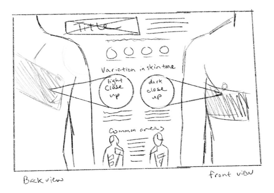
After background research on shingles was conducted, some rough sketches were created to determine the best composition for displaying the relevant information. The second initial sketch was chosen to refine because the client liked how the diagonal stripes resembled the patches of shingles. The revised sketch was changed to a horizontal composition to allow more space for the text and figures on the right-hand side. The diagonal stripes from the initial sketch were changed to a 2D graphic design element that was used to define each area of information.
Initial sketch version 1

Initial sketch version 2

Revised sketch: composition determined
PRODUCTION
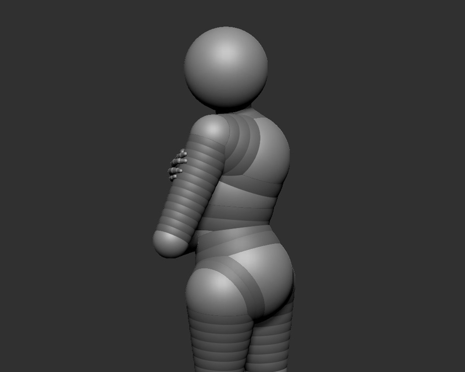
Production of the 2D and 3D assets began once the composition was finalized. The main model of the woman was sculpted in ZBrush, textured in Substance Painter, and rendered in Keyshot. The three circles showing the variations of shingles were created entirely in Substance Painter and rendered in Keyshot. The 2D icons and elements were created in Adobe Illustrator.
Basic proportions and shape of body created using ZSpheres in ZBrush

Low poly model sculpted in ZBrush

Basic shape of head sculpted from a sphere
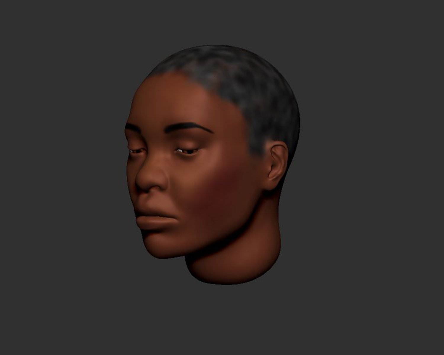
More refined head model

Head was added to the body and more detail was added to the overall model

Materials and texture were added to the model using Substance Painter

Unedited beauty pass render from Keyshot

Final composited image
